REGISTER
LOG IN
HOME
RESOURCES
Publications
Conference Calendar
Past Quiz Results
ARTICLES
Revisiting ISHAM Asia 2021
The first AFWG CaseClinic is now live!
First-ever Study of Mycology Lab Practices in Asia
New Diagnostic Mycology E-learning Course
Antifungal prophylaxis: Whom, what and when
Fereydounia khargensis: A New Opportunistic Yeast Reported from Malaysia
9 Years of MMTN: Improving Fungal Disease Management in Asia Pacific
Echinocandins: Clinicians' Guide
Five controversies in mycology
Fungemia blood culture media
Deep dermatophytosis
AFWG Education Module 4: Is Antifungal Susceptibility Testing Useful for Clinical Management?
AFWG Education Module 5: TDM of Antifungal Agents - Essential or Optional?
AFWG Education Module 6: Antifungal Stewardship
10 common mistakes in laboratory mycology
Itraconazole: A Quick Guide for Clinicians
Evolving Fungal Landscape in Asia
10 common mistakes in clinical mycology
Laboratory Diagnosis of Pythiosis
ICMR Issues C. auris Advisory
Strengths and Limitations of Imaging for Diagnosis of IFI
Candidemia: Lessons Learned from Asian Studies for Intervention
Spotting invasive pulmonary aspergillosis in COVID-19 patients
Pivotal Asian Invasive Mold Study
Impact of the COVID-19 pandemic on IFI epidemiology and trends
Mycetoma in Asia: Still veiled in mystery
Cryptococcosis
Identifying IFI risk factors in patients with COVID-19
ASID ANZMIG x AFWG: Fungal Frontiers in the Asia Pacific – Webinar 2
New Antifungal Agents
Gilead IFI Masterclass: Current updates on the management of IFIs in immunocompromised hosts
The AFWG Masterclass: Advanced fungal education at your fingertips
A challenging case: A crisis unfolds
The role of antifungal stewardship in improving IFI outcomes
Making Precise Diagnoses: Experience from the Laboratory Skills Enhancement Course
A challenging case: A 68-year-old man with nasal and palatal ulcers
AFWG Online Education Module 3: Optimizing Dosing in IFI Management
AFWG Online Education Module 2: Antifungal Prophylaxis in Solid Organ Transplantation
AFWG Education Module 1: The Value of Clinical Mycology Laboratories
How do I interpret Candida in the urine?
How do I interpret Candida in respiratory tract cultures?
Cryptococcosis in HIV and non-HIV infected patients
Human Pythiosis
AFWGOnline Privacy Policy has been Updated
Management of fungal infections in high-risk patients
Striving for Perfection: Experience from the Laboratory Foundation Training Course
Know your fungal landscape in Vietnam
Recent Advances of Fungal Diagnostics in Asian Laboratories
Deep Dermatophytosis: A Case Report
Management of cryptococcosis and talaromycosis
A challenging case: A 49-year-old woman with sarcoidosis
Emerging yeast infections in Asia
Outbreak of Superbug Candida auris: Asian Scenario and Interventions
Championing Medical Mycology: Thoughts on the AFWG Laboratory Skills Enhancement Course
Mucormycosis and Pythiosis – New Insights
AML and the high risk of multiple infectious complications
Do We Need Modification of Recent IDSA & ECIL Guidelines while Managing Patients in Asia?
A hospital’s experience with candidemia and empirical therapy
Top 5 most viewed AFWG videos on YouTube
Fungal Academy 2015
Fluconazole in 2015
Fungal isolation protocol
Influencing Aspergillus
Fungal Asthma
Aspergillus
Laboratory Diagnosis of IPA
Two-Hot-to-Handle
Voriconazole
Educational Organizations
Literature Updates
 

Articles

Cryptococcosis

Share this
page


Share
this page

The pathogenic yeast cell with a polysaccharide capsule, Cryptococcus neoformans/C. gattii complex, causes a life-threatening infection called cryptococcosis. The natural habitats of Cryptococcus are soil with bird (eg, pigeon) droppings, decaying wood and the bark of certain trees. Recently, evidence showed that C. gattii could be isolated from some tropical trees, such as almond trees (Terminalia catappa), pottery trees (Ficus microcarpa) and cassia (Cassia grandis).

Cryptococcosis usually affects immunocompromised hosts, especially HIV patients, via the respiratory tract. After inhalation of either sexual or asexual spores, clinical symptoms develop and manifest in the lungs, blood and, finally, the brain and meninges, sometimes spreading to the lymph nodes and skin. Severe symptoms are seen in both humans and animals. Recently, laboratory surveillance studies have illustrated the variety of Cryptococcus, from the serotype to the molecular level (Table 1).

Table 1. Genus, species, serotypes and molecular types of pathogenic Cryptococcus

Genus and species

Serotype

Molecular type

C. neoformans complex

C. neoformans var. grubii


C. neoformans var. neoformans

A

D

AFLP1/VNI

AFLP1A, AFLP1B/VNII

AFLP2/VNIV

C. gattii complex

C. gattii sensu stricto

C. bacillisporus

C. deuterogattii

C. tetragattii

C. decagattii

B

C

AFLP4/VGI

AFLP5/VGIII

AFLP6/VGII

AFLP7/VGIV

AFLP10/VGIV

HybridsAD, AB, BDAFLP3/VNIII


Worldwide, about 1 million cases of cryptococcal meningitis occur among people with HIV/AIDS each year, resulting in nearly 625,000 deaths. This infection can be found globally, affecting sub-Saharan Africa, USA, Europe and Asia, including Thailand, where the burden of cryptococcal meningitis was estimated at 4.2/100,000 per year.

The majority of these cases in Thailand were in HIV-infected hosts and most of the isolates were C. neoformans var. grubii, serotype A/VNI, which is similar to findings in other countries. A summary of surveillance data from Southeast Asia is shown in Table 2, and includes cultures isolated from 1993 to 2011. Also similar to that seen in other countries, C. gattii complex has been on an upward trend in Southeast Asia from both clinical and environmental sources. A molecular study in Thailand showed a strong link between environmental and clinical strains for the VNI isolates, and similarities among the situations in Thailand and Vancouver (Canada), Australia and South America.

Moreover, a number of unique sequence types have been found in Thailand, which warrants further study in this region. Regarding the susceptibility profile, no resistance strains were presented in these studies, despite reports of resistance from other countries, such as Cambodia. Resistance should be monitored carefully, particularly since non–HIV-infected hosts have been reported.

For brain and meningeal infections, cerebrospinal fluid (CSF) and serum are the preferred specimens for laboratory diagnosis. India ink or nigrosin preparation is used to stain the background for simple direct examination. To increase the yield, one drop of sediment after 1,500 g/3,500 rpm for 10 minutes of centrifugation is recommended for CSF. Sputum, lymph node and tissue biopsy samples can also be used. The supernatant can be used for antigen detection either by lateral flow strip or latex agglutination test. To shorten the identification process, the specimens may be inoculated in the medium with phenoloxidase substrate (caffeic acid/mustard seed/cabbage seed) to induce the melanin production, which is a simple test to confirm the encapsulated yeasts as C. neoformans. Urease production of the encapsulated yeast is another rapid screening test for genus Cryptococcus. Concanavalin medium is one of the recommended tests that can be used to identify the variety. URA5-RFLP, multilocus sequence typing and other molecular techniques are also able to identify the genetic variation for both diagnostic and epidemiologic purposes.
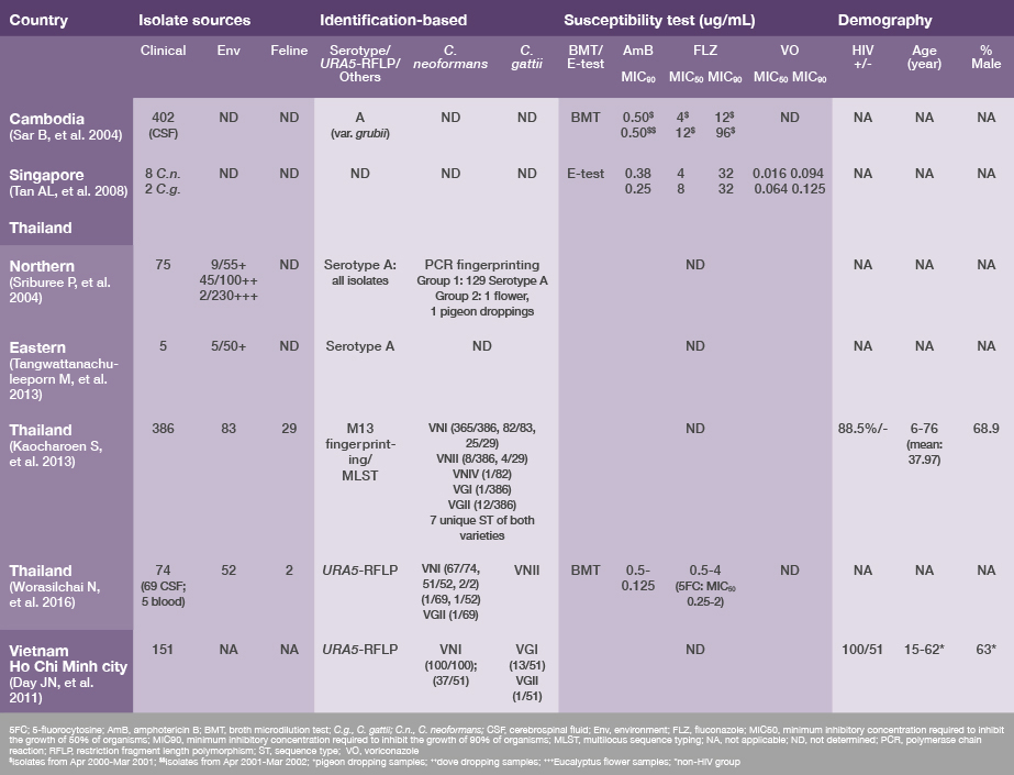
Table 2. Laboratory-based surveillance data on C. neoformans/C. gattii complex in Southeast Asia

table2-lab-surveillance-data-cneoformans-cgattii-sea

References:

  1. Chayakulkeeree M, Denning DW. LIFE website. Available at: www.life-worldwide.org/assets/uploads/files/Thailand.pdf.
  2. Day JN, et al. J Clin Microbiol 2011;49:658-664.
  3. Desnos-Ollivier M, et al. MBio 2015;6:e00311.
  4. Hagen F, et al. Fungal Genet Biol 2015;78:16-48.
  5. Kaocharoen S, et al. PLoS Negl Trop Dis 2013;7:e2297.
  6. Sar B, et al. J Antimicrob Chemother 2004;54:563-565.
  7. Sriburee P, et al. Mycopathologia 2004;158:25-31.
  8. Tan AL, Chan KS. Ann Acad Med Singapore 2008;37:841-846.
  9. Tangwattanachuleeporn M, et al. Med Mycol J 2013;54:303-307.
  10. Worasilchai N, et al. Med Mycol 2016 (in press).
SIGN UP FOR NEWSLETTER
SIGN UP

This field is required. Please enter your email address.
Thank you for signing up for the AFWG newsletter.
Your subscription has been updated.